 Fraunhofer Institute for Mechanics of Materials IWM
Microstructure analysis via EBSD
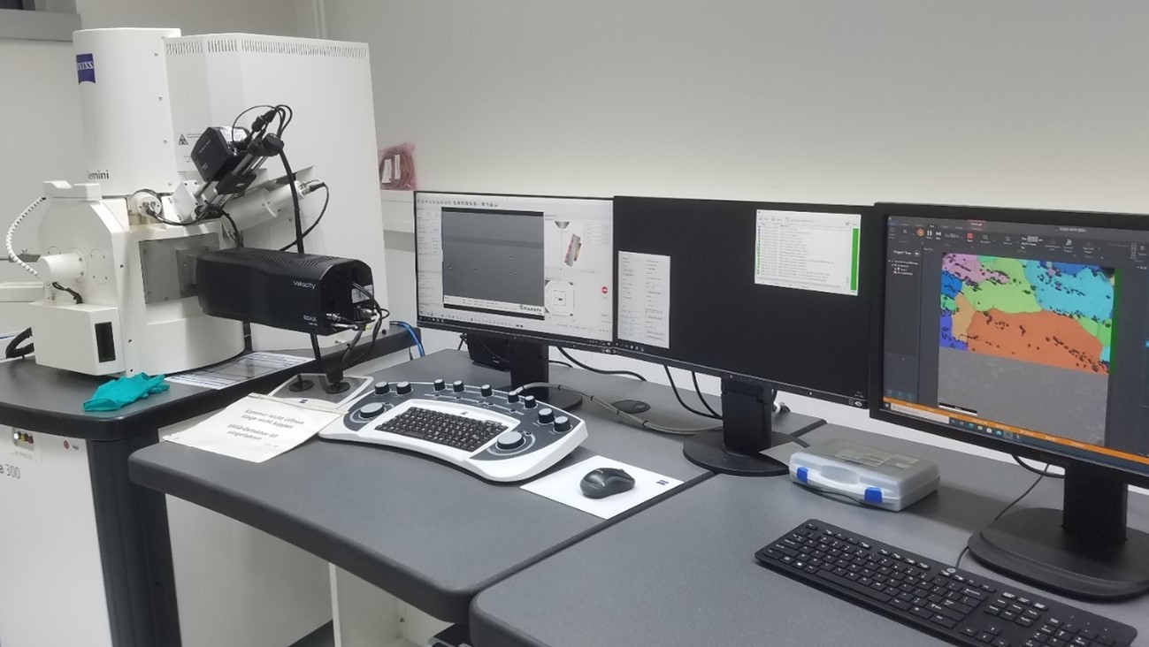
Fig. 1: Field emission scanning electron microscope from Zeiss with EBSD and EDX system from EDAX
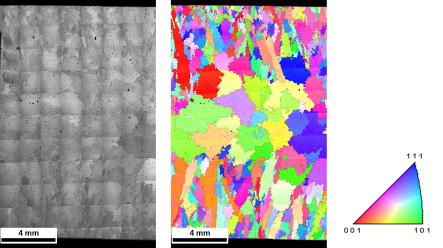
Fig. 2: Coarse-grained cast structure of a nickel-based alloy. An EBSD assembly scan was used to scan an area of approx. 12mm x 18mm over the entire sample thickness.
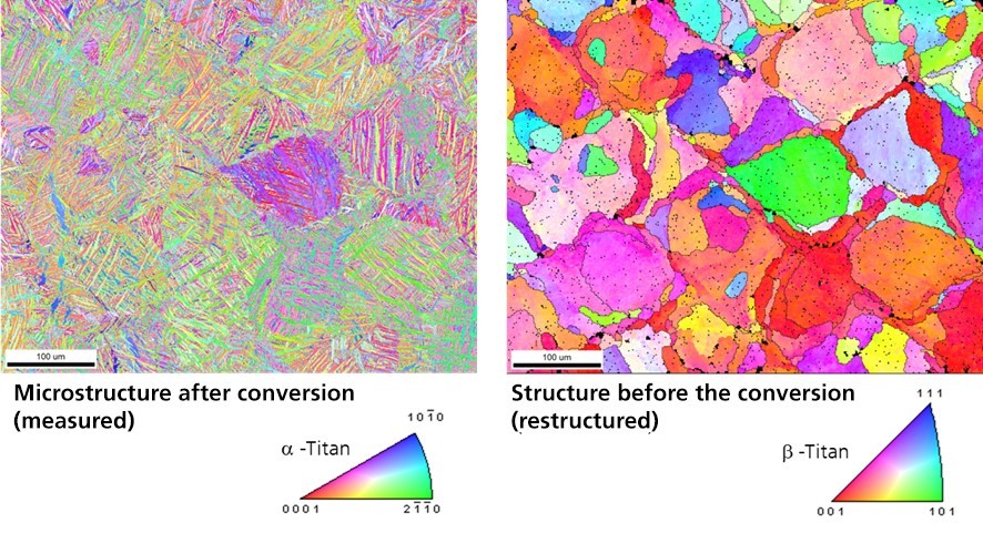
Fig. 3: EBSD image of an additively manufactured sample made of Ti-6Al-4V: The crystal orientations are shown in color, on the left of the martensitic microstructure of the hexagonal α-phase and on the right of the reconstructed microstructure of the body-centered cubic high-temperature phase β including the reconstructed grain boundaries. The reconstruction was based on Burgers' orientation relationship ({110}ᵦ|| {0001}α and <1-11>ᵦ || <11-20>α with OIM AnalysisTM 8.6.
The EBSD (Electron Backscatter Diffraction) method is based on the diffraction of electrons at lattice planes of crystalline samples, analogous to X-ray or neutron diffraction (Bragg's equation: 2dsinθ=nλ) with the special feature that EBSD is a spatially resolved method. The crystallographic orientation is determined for each measuring point, from which many microstructural parameters can be quantified and visualized.
The Sigma 300 scanning electron microscope from Zeiss acquired by the Fraunhofer IWM in 2021 is equipped with a high-speed EBSD camera from EDAX (Fig. 1). Thanks to short acquisition times, high computing power and a sizable capacity for storage, large measuring ranges with high resolution can now be realized. It is also possible to create assembly scans: a defined measuring area is scanned by motorized table control in the SEM and the individual scans are automatically assembled. Fig. 2 shows a very coarse-grained cast structure of a nickel-based alloy. Large scan areas are necessary to be able to display the grains in the center of the sample with diameters of several millimeters. Another example shown in Fig. 3 is an EBSD analysis of additively manufactured Ti-6Al-4V. The resulting microstructure is martensitic (α-titanium). The reconstruction of the initial grain structure that was present at high temperature during the manufacturing process is usually possible with Ti alloys, based on their limited number as well as the precisely defined orientation relationships between the forming α-phase and the high-temperature β-phase. With the aid of a reconstruction, the microstructure before the phase transformation can in many cases therefore be reconstructed from the EBSD scans of transformation structures (Fig. 3).